Locally Modulating T cells to Treat Systemic Inflammation
Experience Synopsis: I was the lead researcher responsible for developing a novel therapeutic agent to locally modulate T cells in inflammatory arthritis affected joints. After making and testing a series of formulations, I demonstrated that it was possible to use a biomaterial to turn a single joint into a “factory” to produce inflammation resolving immune cells that recirculate throughout the body to resolve joint-associated inflammation without broadly immunosuppressive side-effects.
Key Contributions and Takeaways

Experimental Design
Designed, implemented and analyzed experiments to characterize formulations and determine immunological mechanism of action using a variety of techniques including flow cytometry, next-gen sequencing, qPCR, ELISA and more.

Funding Acquisiton
I was a key contributor on all grants related to the project, outlining research goals, preparing preliminary data sections, and providing experimental design to reach research goals. The project resulted in the acquisition of ~$5 M in funding from various sources.

Focus on Translation
During the project, I focused on developing the therapy in a manner to allow for clinical translation. The therapy is continuing to be developed in a spinout company, for which I assisted with the business plan design, market analysis, customer discovery interviews, regulatory and legal strategy, and IND-enabling study design.
Skills Used

Flow Cytometry
Multicolor flow cytometry was used to assess phenotypic fates of inflammation-associated immune cell populations including T cells, macrophages, and dendritic cells.

Epigenetic Sequencing
ATAC sequencing, CUT-and-TAG sequencing, and Reduced Representation Bisulfite Sequencing were used to assess epigenetic changes in cells.

In Vivo Modeling
Multiple inflammatory arthritis models were used to assess the efficacy of the treatment and provide insights into the mechanism of action.

Cell Culture
Murine and human primary cell culture was used to perform ex vivo assays to determine the impact of active compounds on cell phenotypes.

Histology and Micro-CT
Histology and micro-computed tomography (CAT scans) were used to assess inflammation, bone erosion and disease progression.

Scanning Electron Microscopy
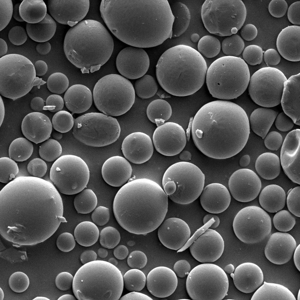
Scanning electron microscopy was used to characterized material formulations and morphological properties.